| UTHEALTH HOME ABOUT SBMI A-Z WEBMAIL INSIDE THE UNIVERSITY |
|
|||||||
|

Kinase Fusion Gene:LMTK2_SRPK2 |
Kinase Fusion Protein Summary |
 Kinase Fusion gene summary Kinase Fusion gene summary |
| Kinase Fusion partner gene information | Kinase Fusion gene name: LMTK2_SRPK2 | KinaseFusionDB ID: KFG3190 | FusionGDB2.0 ID: KFG3190 | Hgene | Tgene | Gene symbol | LMTK2 | SRPK2 | Gene ID | 22853 | 6733 | |
| Gene name | lemur tyrosine kinase 2 | SRSF protein kinase 2 | ||||||||||
| Synonyms | AATYK2|BREK|KPI-2|KPI2|LMR2|PPP1R100|cprk|hBREK | SFRSK2 | ||||||||||
| Cytomap | 7q21.3 | 7q22.3 | ||||||||||
| Type of gene | protein-coding | protein-coding | ||||||||||
| Description | serine/threonine-protein kinase LMTK2CDK5/p35-regulated kinaseapoptosis-associated tyrosine kinase 2brain-enriched kinasecyclin-dependent kinase 5/p35-regulated kinasekinase/phosphatase/inhibitor 2protein phosphatase 1, regulatory subunit 100serine | SRSF protein kinase 2SFRS protein kinase 2SR protein kinase 2SR-protein-specific kinase 2serine kinase SRPK2serine/arginine-rich protein-specific kinase 2serine/arginine-rich splicing factor kinase 2serine/threonine-protein kinase SRPK2 | ||||||||||
| Modification date | 20240411 | 20240403 | ||||||||||
| UniProtAcc | Q8IWU2 | P78362 | ||||||||||
| Ensembl transtripts involved in fusion gene | ENST ids | ENST00000297293, ENST00000493372, | ENST00000493638, ENST00000357311, ENST00000393651, ENST00000489828, | |||||||||
| Context (manual curation of fusion genes in KinaseFusionDB) | PubMed: LMTK2 [Title/Abstract] AND SRPK2 [Title/Abstract] AND fusion [Title/Abstract] | |||||||||||
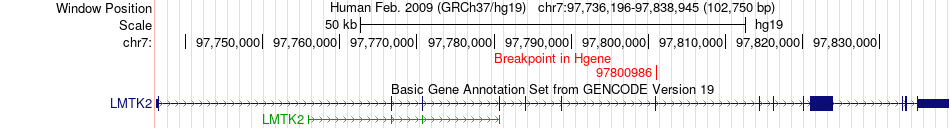
| Most frequent breakpoint (based on all fusion genes of FusionGDB 2.0) | LMTK2(97800986)-SRPK2(104844232), # samples:1 | |||||||||||
| Gene ontology of each fusion partner gene with evidence of Inferred from Direct Assay (IDA) from Entrez |
| Partner | Gene | GO ID | GO term | PubMed ID |
| Hgene | LMTK2 | GO:0006468 | protein phosphorylation | 12393858 |
| Hgene | LMTK2 | GO:0018105 | peptidyl-serine phosphorylation | 16887929 |
| Hgene | LMTK2 | GO:0018107 | peptidyl-threonine phosphorylation | 16887929 |
| Hgene | LMTK2 | GO:0046777 | protein autophosphorylation | 12393858 |
| Tgene | SRPK2 | GO:0000245 | spliceosomal complex assembly | 9472028 |
| Tgene | SRPK2 | GO:0006468 | protein phosphorylation | 9472028 |
| Tgene | SRPK2 | GO:0008284 | positive regulation of cell population proliferation | 18559500 |
| Tgene | SRPK2 | GO:0008380 | RNA splicing | 9472028 |
| Tgene | SRPK2 | GO:0035556 | intracellular signal transduction | 9472028 |
| Tgene | SRPK2 | GO:0045070 | positive regulation of viral genome replication | 20498328 |
| Tgene | SRPK2 | GO:0045071 | negative regulation of viral genome replication | 12417631 |
| Tgene | SRPK2 | GO:0062176 | R-loop processing | 28076779 |
| Kinase Fusion gene breakpoints across LMTK2 (5'-gene) * Click on the image to open the UCSC genome browser with custom track showing this image in a new window. |
 |
| Kinase Fusion gene breakpoints across SRPK2 (3'-gene) * Click on the image to open the UCSC genome browser with custom track showing this image in a new window. |
 |
Top |
Kinase Fusion Gene Sample Information |
| Kinase Fusion gene information. |
| Kinase Fusion gene information from four resources (ChiTars 5.0, ChimerDB 4.0, COSMIC, and CCLE) * All genome coordinats were lifted-over on hg19. * Click on the break point to see the gene structure around the break point region using the UCSC Genome Browser. |
| Source | Sample | Hgene | Hchr | Hbp | Tgene | Tchr | Tbp |
| ChimerDB4 | 5263N | LMTK2 | chr7 | 97800986 | SRPK2 | chr7 | 104844232 |
Top |
Kinase Fusion ORF Analysis |
| Kinase Fusion information from ORFfinder translation from full-length transcript sequence from KinaseFusionDB. |
| Henst | Tenst | Hgene | Hchr | Hbp | Tgene | Tchr | Tbp | Seq length (transcript) | Seq length (amino acids) |
| ENST00000297293 | ENST00000393651 | LMTK2 | chr7 | 97800986 | SRPK2 | chr7 | 104844232 | 4575 | 939 |
Top |
Kinase Fusion Amino Acid Sequences |
| For individual full-length fusion transcript sequence from KinaseFusionDB, we ran ORFfinder and chose the longest ORF among the all predicted ones. |
| >Henst_Tenst_Hgene_Hchr_Hbp_Tgene_Tchr_Tbp_length(fusion AA)_AAseq >ENST00000297293_ENST00000393651_LMTK2_chr7_97800986_SRPK2_chr7_104844232_length(amino acids)=939 MPGPPALRRRLLLLLLVLLIAGSAGAAPLPQTGAGEAPPAAEVSSSFVILCVCSLIILIVLIANCVSCCKDPEIDFKEFEDNFDDEIDFT PPAEDTPSVQSPAEVFTLSVPNISLPAPSQFQPSVEGLKSQVARHSLNYIQEIGNGWFGKVLLGEIYTGTSVARVIVKELKASANPKEQD TFLKNGEPYYILQHPNILQCVGQCVEAIPYLLVFEFCDLGDLKAYLRSEQEHMRGDSQTMLLQRMACEVAAGLAAMHKLHFLHRPEPQQK APLVPPPPPPPPPPPPPLPDPTPPEPEEEILGSDDEEQEDPADYCKGGYHPVKIGDLFNGRYHVIRKLGWGHFSTVWLCWDMQGKRFVAM KVVKSAQHYTETALDEIKLLKCVRESDPSDPNKDMVVQLIDDFKISGMNGIHVCMVFEVLGHHLLKWIIKSNYQGLPVRCVKSIIRQVLQ GLDYLHSKCKIIHTDIKPENILMCVDDAYVRRMAAEATEWQKAGAPPPSGSAVSTAPQQKPIGKISKNKKKKLKKKQKRQAELLEKRLQE IEELEREAERKIIEENITSAAPSNDQDGEYCPEVKLKTTGLEEAAEAETAKDNGEAEDQEEKEDAEKENIEKDEDDVDQELANIDPTWIE SPKTNGHIENGPFSLEQQLDDEDDDEEDCPNPEEYNLDEPNAESDYTYSSSYEQFNGELPNGRHKIPESQFPEFSTSLFSGSLEPVACGS VLSEGSPLTEQEESSPSHDRSRTVSASSTGDLPKAKTRAADLLVNPLDPRNADKIRVKIADLGNACWVHKHFTEDIQTRQYRSIEVLIGA GYSTPADIWSTACMAFELATGDYLFEPHSGEDYSRDEDHIAHIIELLGSIPRHFALSGKYSREFFNRRGELRHITKLKPWSLFDVLVEKY -------------------------------------------------------------- |
Multiple Sequence Alignment of All Fusion Protein Isoforms |
Top |
Kinase Fusion Protein Functional Features |
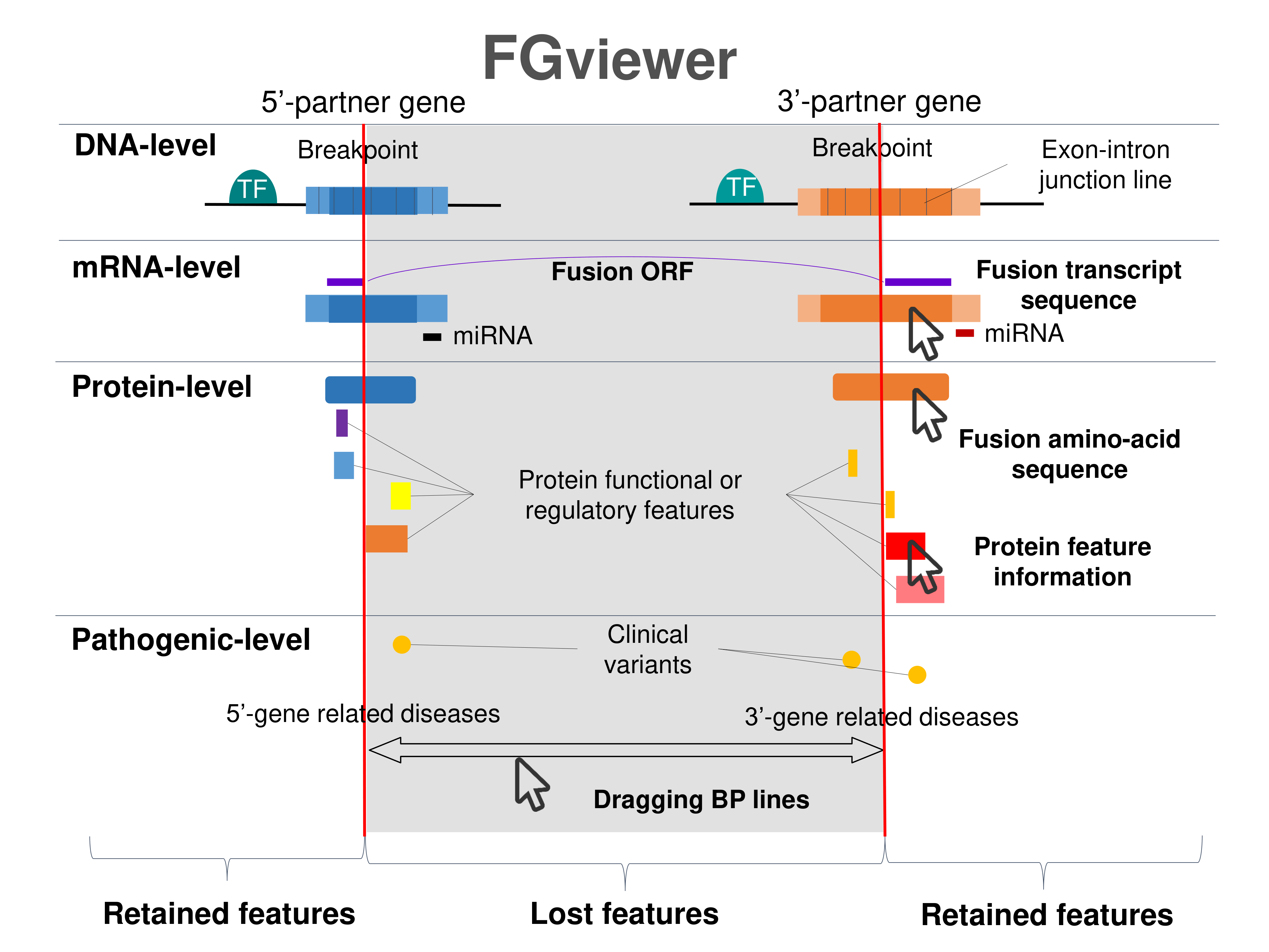
| Four levels of functional features of fusion genes Go to FGviewer search page for the most frequent breakpoint (https://ccsmweb.uth.edu/FGviewer/chr7:97800986/chr7:104844232) - FGviewer provides the online visualization of the retention search of the protein functional features across DNA, RNA, protein, and pathological levels. - How to search 1. Put your fusion gene symbol. 2. Press the tab key until there will be shown the breakpoint information filled. 4. Go down and press 'Search' tab twice. 4. Go down to have the hyperlink of the search result. 5. Click the hyperlink. 6. See the FGviewer result for your fusion gene. |
 |
| Main function of each fusion partner protein. (from UniProt) |
| Hgene | Tgene |
| LMTK2 | SRPK2 |
| FUNCTION: Phosphorylates PPP1C, phosphorylase b and CFTR. | FUNCTION: Serine/arginine-rich protein-specific kinase which specifically phosphorylates its substrates at serine residues located in regions rich in arginine/serine dipeptides, known as RS domains and is involved in the phosphorylation of SR splicing factors and the regulation of splicing (PubMed:9472028, PubMed:18559500, PubMed:21056976). Promotes neuronal apoptosis by up-regulating cyclin-D1 (CCND1) expression (PubMed:19592491). This is done by the phosphorylation of SRSF2, leading to the suppression of p53/TP53 phosphorylation thereby relieving the repressive effect of p53/TP53 on cyclin-D1 (CCND1) expression (PubMed:21205200). Phosphorylates ACIN1, and redistributes it from the nuclear speckles to the nucleoplasm, resulting in cyclin A1 but not cyclin A2 up-regulation (PubMed:18559500). Plays an essential role in spliceosomal B complex formation via the phosphorylation of DDX23/PRP28 (PubMed:18425142). Probably by phosphorylating DDX23, leads to the suppression of incorrect R-loops formed during transcription; R-loops are composed of a DNA:RNA hybrid and the associated non-template single-stranded DNA (PubMed:28076779). Can mediate hepatitis B virus (HBV) core protein phosphorylation (PubMed:12134018). Plays a negative role in the regulation of HBV replication through a mechanism not involving the phosphorylation of the core protein but by reducing the packaging efficiency of the pregenomic RNA (pgRNA) without affecting the formation of the viral core particles (PubMed:16122776). {ECO:0000269|PubMed:12134018, ECO:0000269|PubMed:16122776, ECO:0000269|PubMed:18425142, ECO:0000269|PubMed:18559500, ECO:0000269|PubMed:19592491, ECO:0000269|PubMed:21056976, ECO:0000269|PubMed:21205200, ECO:0000269|PubMed:28076779, ECO:0000269|PubMed:9472028}. |
| Retention analysis result of each fusion partner protein across 39 protein features of UniProt such as six molecule processing features, 13 region features, four site features, six amino acid modification features, two natural variation features, five experimental info features, and 3 secondary structure features. Here, because of limited space for viewing, we only show the protein feature retention information belong to the 13 regional features. All retention annotation result can be downloaded at * Minus value of BPloci means that the break pointn is located before the CDS. |
| - Retained domain in the 5'-partner of fusion protein (protein functional feature from UniProt). |
| Partner | Hgeneene | Hbp | Tgeneene | Tbp | ENST | BPexon | TotalExon | Protein feature loci | BPloci | TotalLen | Feature | Note |
| - Retained domain in the 3'-partner of fusion protein (protein functional feature from UniProt). |
| Partner | Hgeneene | Hbp | Tgeneene | Tbp | ENST | BPexon | TotalExon | Protein feature loci | BPloci | TotalLen | Feature | Note |
| Tgene | LMTK2 | 97800986 | SRPK2 | 104844232 | ENST00000297293 | 0 | 15 | 81_684 | 12 | 689 | Domain | Note=Protein kinase;Ontology_term=ECO:0000255;evidence=ECO:0000255|PROSITE-ProRule:PRU00159 |
| Tgene | LMTK2 | 97800986 | SRPK2 | 104844232 | ENST00000297293 | 1 | 16 | 81_684 | 23 | 700 | Domain | Note=Protein kinase;Ontology_term=ECO:0000255;evidence=ECO:0000255|PROSITE-ProRule:PRU00159 |
| Tgene | LMTK2 | 97800986 | SRPK2 | 104844232 | ENST00000297293 | 2 | 17 | 81_684 | 12 | 689 | Domain | Note=Protein kinase;Ontology_term=ECO:0000255;evidence=ECO:0000255|PROSITE-ProRule:PRU00159 |
Top |
Kinase Fusion Protein Structures |
| CIF files of the predicted kinase fusion proteins * Here we show the 3D structure of the fusion proteins using Mol*. AlphaFold produces a per-residue confidence score (pLDDT) between 0 and 100. Model confidence is shown from the pLDDT values per residue. pLDDT corresponds to the model’s prediction of its score on the local Distance Difference Test. It is a measure of local accuracy (from AlphfaFold website). To color code individual residues, we transformed individual PDB files into CIF format. |
| Kinase Fusion protein CIF link (fusion AA seq ID in KinaseFusionDB) | Henst | Tenst | Hgene | Hchr | Hbp | Tgene | Tchr | Tbp | AA seq | Len(AA seq) |
| PDB file >>>285_LMTK2_SRPK2 | ENST00000297293 | ENST00000393651 | LMTK2 | chr7 | 97800986 | SRPK2 | chr7 | 104844232 | MPGPPALRRRLLLLLLVLLIAGSAGAAPLPQTGAGEAPPAAEVSSSFVILCVCSLIILIVLIANCVSCCKDPEIDFKEFEDNFDDEIDFT PPAEDTPSVQSPAEVFTLSVPNISLPAPSQFQPSVEGLKSQVARHSLNYIQEIGNGWFGKVLLGEIYTGTSVARVIVKELKASANPKEQD TFLKNGEPYYILQHPNILQCVGQCVEAIPYLLVFEFCDLGDLKAYLRSEQEHMRGDSQTMLLQRMACEVAAGLAAMHKLHFLHRPEPQQK APLVPPPPPPPPPPPPPLPDPTPPEPEEEILGSDDEEQEDPADYCKGGYHPVKIGDLFNGRYHVIRKLGWGHFSTVWLCWDMQGKRFVAM KVVKSAQHYTETALDEIKLLKCVRESDPSDPNKDMVVQLIDDFKISGMNGIHVCMVFEVLGHHLLKWIIKSNYQGLPVRCVKSIIRQVLQ GLDYLHSKCKIIHTDIKPENILMCVDDAYVRRMAAEATEWQKAGAPPPSGSAVSTAPQQKPIGKISKNKKKKLKKKQKRQAELLEKRLQE IEELEREAERKIIEENITSAAPSNDQDGEYCPEVKLKTTGLEEAAEAETAKDNGEAEDQEEKEDAEKENIEKDEDDVDQELANIDPTWIE SPKTNGHIENGPFSLEQQLDDEDDDEEDCPNPEEYNLDEPNAESDYTYSSSYEQFNGELPNGRHKIPESQFPEFSTSLFSGSLEPVACGS VLSEGSPLTEQEESSPSHDRSRTVSASSTGDLPKAKTRAADLLVNPLDPRNADKIRVKIADLGNACWVHKHFTEDIQTRQYRSIEVLIGA GYSTPADIWSTACMAFELATGDYLFEPHSGEDYSRDEDHIAHIIELLGSIPRHFALSGKYSREFFNRRGELRHITKLKPWSLFDVLVEKY | 939 |
| 3D view using mol* of 285_LMTK2_SRPK2 | ||||||||||
| PDB file >>>TKFP_499_LMTK2_SRPK2 | ENST00000297293 | ENST00000393651 | LMTK2 | chr7 | 97800986 | SRPK2 | chr7 | 104844232 | MPGPPALRRRLLLLLLVLLIAGSAGAAPLPQTGAGEAPPAAEVSSSFVILCVCSLIILIVLIANCVSCCKDPEIDFKEFEDNFDDEIDFT PPAEDTPSVQSPAEVFTLSVPNISLPAPSQFQPSVEGLKSQVARHSLNYIQEIGNGWFGKVLLGEIYTGTSVARVIVKELKASANPKEQD TFLKNGEPYYILQHPNILQCVGQCVEAIPYLLVFEFCDLGDLKAYLRSEQEHMRGDSQTMLLQRMACEVAAGLAAMHKLHFLHRPEPQQK APLVPPPPPPPPPPPPPLPDPTPPEPEEEILGSDDEEQEDPADYCKGGYHPVKIGDLFNGRYHVIRKLGWGHFSTVWLCWDMQGKRFVAM KVVKSAQHYTETALDEIKLLKCVRESDPSDPNKDMVVQLIDDFKISGMNGIHVCMVFEVLGHHLLKWIIKSNYQGLPVRCVKSIIRQVLQ GLDYLHSKCKIIHTDIKPENILMCVDDAYVRRMAAEATEWQKAGAPPPSGSAVSTAPQQKPIGKISKNKKKKLKKKQKRQAELLEKRLQE IEELEREAERKIIEENITSAAPSNDQDGEYCPEVKLKTTGLEEAAEAETAKDNGEAEDQEEKEDAEKENIEKDEDDVDQELANIDPTWIE SPKTNGHIENGPFSLEQQLDDEDDDEEDCPNPEEYNLDEPNAESDYTYSSSYEQFNGELPNGRHKIPESQFPEFSTSLFSGSLEPVACGS VLSEGSPLTEQEESSPSHDRSRTVSASSTGDLPKAKTRAADLLVNPLDPRNADKIRVKIADLGNACWVHKHFTEDIQTRQYRSIEVLIGA GYSTPADIWSTACMAFELATGDYLFEPHSGEDYSRDEDHIAHIIELLGSIPRHFALSGKYSREFFNRRGELRHITKLKPWSLFDVLVEKY | 939_LMTK2_SRPK2 |
Top |
Comparison of Fusion Protein Isoforms |
| Superimpose the 3D Structures Among All Fusion Protein Isoforms * Download the pdb file and open it from the molstar online viewer. |
| Comparison of the Secondary Structures of Fusion Protein Isoforms |
Top |
Comparison of Fusion Protein Sequences/Structures with Known Sequences/Structures from PDB |
Top |
pLDDT score distribution |
| pLDDT score distribution of the predicted fusion protein structures from AlphaFold2 * AlphaFold produces a per-residue confidence score (pLDDT) between 0 and 100. * The blue color at the bottom marks the best active site residues. |
| 285_LMTK2_SRPK2.png |
 |
| 285_LMTK2_SRPK2.png |
 |
Top |
Potential Active Site Information |
| The potential binding sites of these fusion proteins were identified using SiteMap, a module of the Schrodinger suite. |
| Kinase Fusion AA seq ID in KinaseFusionDB | Site score | Size | Dscore | Volume | Exposure | Enclosure | Contact | Phobic | Philic | Balance | Don/Acc | Residues |
Top |
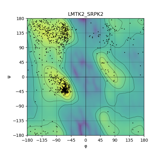
Ramachandran Plot of Kinase Fusion Protein Structure |
| Ramachandran plot of the torsional angles - phi (φ)and psi (ψ) - of the residues (amino acids) contained in this fusion protein peptide. |
| 285_LMTK2_SRPK2_ramachandran.png |
 |
Top |
Virtual Screening Results |
| Distribution of the average docking score across all approved kinase inhibitors. Distribution of the number of occurrence across all approved kinase inhibitors. |
| 5'-kinase fusion protein case |
| 3'-kinase fusion protein case |
Top |
| Drug information from DrugBank of the top 20 interacting small molecules. * The detailed information of individual kinase inhibitors are available in the download page. |
| Fusion gene name info | Drug | Docking score | Glide g score | Glide energy |
Top |
Kinase-Substrate Information of LMTK2_SRPK2 |
| Phosphorylation target of the kinase (phosphosite, 03-17-2024) |
| Kinase | Kinase UniProt Acc | Kinase species | Substrate | Substrate UniProt Acc | Substrate phosphorylated residues | Substrate phosphorylated sites (+/-7AA) | Domain |
| LMTK2 | Q8IWU2 | human | PPP1CA | P62136 | T320 | NPGGRPItPPRNsAK | |
| LMTK2 | Q8IWU2 | human | CFTR | P13569 | S737 | EPLERRLsLVPDSEQ | CFTR_R |
| LMTK2 | Q8IWU2 | human | PYGB | P11216 | S15 | sEkRKQIsVRGLAGL | |
| SRPK2 | P78362 | human | LGMN | Q99538 | S226 | CYYDEkRsTYLGDWY | Peptidase_C13 |
| SRPK2 | P78362 | human | ACIN1 | Q9UKV3 | S1180 | GPRsRsRsRDRRRKE | RSB_motif |
| SRPK2 | P78362 | human | SRPK2 | P78362 | S494 | HDRSRtVsAsstGDL | |
| SRPK2 | P78362 | human | MAPT | P10636 | S214 | GGKERPGsKEEVDED |
| Biological Network Integration of This Kinase and Substrates (GeneMANIA website) |
| Enriched GO biological processes of the phosphorylation target genes of the kinase |
| Kinase | GOID | GO term | P.adjust |
| LMTK2 | ID | Description | 0.00e+00 |
| LMTK2 | GO:0005980 | glycogen catabolic process | 2.26e-04 |
| LMTK2 | GO:0009251 | glucan catabolic process | 2.26e-04 |
| LMTK2 | GO:0000272 | polysaccharide catabolic process | 2.26e-04 |
| LMTK2 | GO:0005977 | glycogen metabolic process | 1.94e-03 |
| LMTK2 | GO:0044042 | glucan metabolic process | 1.94e-03 |
| LMTK2 | GO:0006112 | energy reserve metabolic process | 2.11e-03 |
| LMTK2 | GO:0005976 | polysaccharide metabolic process | 2.37e-03 |
| LMTK2 | GO:0016052 | carbohydrate catabolic process | 5.31e-03 |
| LMTK2 | GO:0015980 | energy derivation by oxidation of organic compounds | 2.01e-02 |
| LMTK2 | GO:0042045 | epithelial fluid transport | 2.51e-02 |
| LMTK2 | GO:0060081 | membrane hyperpolarization | 2.51e-02 |
| LMTK2 | GO:2001241 | positive regulation of extrinsic apoptotic signaling pathway in absence of ligand | 2.51e-02 |
| LMTK2 | GO:0070170 | regulation of tooth mineralization | 2.51e-02 |
| LMTK2 | GO:0043558 | regulation of translational initiation in response to stress | 2.51e-02 |
| LMTK2 | GO:0070262 | peptidyl-serine dephosphorylation | 2.51e-02 |
| LMTK2 | GO:1904321 | response to forskolin | 2.51e-02 |
| LMTK2 | GO:1904322 | cellular response to forskolin | 2.51e-02 |
| LMTK2 | GO:0035970 | peptidyl-threonine dephosphorylation | 2.89e-02 |
| LMTK2 | GO:0044070 | regulation of monoatomic anion transport | 2.89e-02 |
| LMTK2 | GO:0010288 | response to lead ion | 2.91e-02 |
| LMTK2 | GO:0043555 | regulation of translation in response to stress | 2.91e-02 |
| LMTK2 | GO:0070166 | enamel mineralization | 2.91e-02 |
| LMTK2 | GO:0043153 | entrainment of circadian clock by photoperiod | 2.95e-02 |
| LMTK2 | GO:0097186 | amelogenesis | 2.95e-02 |
| LMTK2 | GO:0009648 | photoperiodism | 2.95e-02 |
| LMTK2 | GO:0006833 | water transport | 2.95e-02 |
| LMTK2 | GO:0009649 | entrainment of circadian clock | 2.95e-02 |
| LMTK2 | GO:0035774 | positive regulation of insulin secretion involved in cellular response to glucose stimulus | 2.95e-02 |
| LMTK2 | GO:0005979 | regulation of glycogen biosynthetic process | 2.95e-02 |
| LMTK2 | GO:0010962 | regulation of glucan biosynthetic process | 2.95e-02 |
| LMTK2 | GO:0034505 | tooth mineralization | 2.95e-02 |
| LMTK2 | GO:0048240 | sperm capacitation | 2.95e-02 |
| LMTK2 | GO:0050891 | multicellular organismal-level water homeostasis | 3.05e-02 |
| LMTK2 | GO:0015701 | bicarbonate transport | 3.05e-02 |
| LMTK2 | GO:0070633 | transepithelial transport | 3.05e-02 |
| LMTK2 | GO:0042044 | fluid transport | 3.13e-02 |
| LMTK2 | GO:0070873 | regulation of glycogen metabolic process | 3.13e-02 |
| LMTK2 | GO:0032885 | regulation of polysaccharide biosynthetic process | 3.13e-02 |
| LMTK2 | GO:0006904 | vesicle docking involved in exocytosis | 3.26e-02 |
| LMTK2 | GO:0005978 | glycogen biosynthetic process | 3.26e-02 |
| LMTK2 | GO:0009250 | glucan biosynthetic process | 3.26e-02 |
| LMTK2 | GO:0032881 | regulation of polysaccharide metabolic process | 3.26e-02 |
| LMTK2 | GO:2001239 | regulation of extrinsic apoptotic signaling pathway in absence of ligand | 3.26e-02 |
| LMTK2 | GO:0071320 | cellular response to cAMP | 3.46e-02 |
| LMTK2 | GO:2001238 | positive regulation of extrinsic apoptotic signaling pathway | 3.50e-02 |
| LMTK2 | GO:0061178 | regulation of insulin secretion involved in cellular response to glucose stimulus | 3.50e-02 |
| LMTK2 | GO:0070169 | positive regulation of biomineral tissue development | 3.50e-02 |
| LMTK2 | GO:0006695 | cholesterol biosynthetic process | 3.58e-02 |
| LMTK2 | GO:1902653 | secondary alcohol biosynthetic process | 3.58e-02 |
Top |
Related Drugs to LMTK2_SRPK2 |
| Drugs used for this fusion-positive patient. (Manual curation of PubMed, 04-30-2022 + MyCancerGenome) |
| Hgene | Tgene | Drug | Source | PMID |
| Distribution of the number of studies mentioning LMTK2-SRPK2 and kinase inhibitors the PubMed Abstract (04-01-2024) |
| Fusion gene - drug pair 1 | Fusion gene - drug pair 2 | PMID | Publication date | DOI | Study title |
Top |
Related Diseases to LMTK2_SRPK2 |
| Diseases that have this fusion gene. (Manual curation of PubMed, 04-30-2022 + MyCancerGenome) |
| Hgene | Tgene | Disease | Source | PMID |
| Related diseases from the literature mentioned this fusion gene and drug. (PubMed, 04-01-2024) |
| MeSH ID | MeSH term |
| Diseases associated with fusion partners. (DisGeNet 4.0) |
| Partner | Gene | Disease ID | Disease name | # pubmeds | Source |
Top |
Clinical Trials of the Found Drugs/Small Molecules |
| Statistics of the Clinical Trials of the Found Kinase Inibitors from clinicaltrials.gov (06-17-2024) |
| Clinical Trials from clinicaltrials.gov (06-17-2024) |
| Fusion Gene | Kinase Inhibitor | NCT ID | Study Status | Phases | Disease | # Enrolment | Date |
 Copyright 2024-Present -The University of Texas Health Science Center at Houston
Web File Viewing | Emergency Information |Campus Carry|Site Policies |